Diagnostic Centre
Book consultationBoneham Optometrist Technology
At Boneham Optometrist, we continually invest in technology so we can remain at the cutting edge of eye care.
We have innovative technology that is used to detect eye diseases earlier, aid in treatment and enable accurate monitoring of progression.
We have instruments that measure the cornea and sclera, allowing us to design customised contact lenses for conditions such as keratoconus, severe dry eye, pellucid marginal degeneration and progressive myopia. Read about the OPTOS, Topographer, OCT and SMAP below.
Optos Daytona Plus
The retina is the name given to the light sensitive tissue at the back of the eye. As the eye is very curved, standard cameras have been limited to photographing just the central retina, which has the optic nerve and macula. Previous cameras could only capture 40 degrees of the central retina, the Optos allows us to look right out to the edge of the eye and allows a detailed view of the peripheral retina. It covers 5 times the area in one image compared to a regular retinal camera.
This has allowed us to see many conditions that could have been undetected or delayed detection. For example, retinal tears and detachments on the back of the eye, which, in certain circumstances can be quite hard to identify without dilation. These can be easily seen with the Optos. The machine has led to many cases of early detection and interventions of sight threatening conditions.


Corneal Topography
The cornea is the clear tissue at the front of the eye, the role of the cornea is to focus a rays of light onto the retina. If the shape of the cornea isn’t a sphere you have what is known as astigmatism.
The corneal topographer allows us to map the front surface of the cornea and measure astigmatism. It also allows us to detect conditions such as keratoconus and pellucid marginal degeneration. We also use the topographer to guide contact lens design for eye disease and for orthokeratology. We have just upgraded our Medmont topographer to the latest version. The new Meridia topographer not only gives us more information on the corneal shape, allowing us to better fit contact lenses on for example , our keratoconus patients , it also allows us to better diagnose and manage dry eye problems.


Optical Coherence Tomography (OCT)
OCT is used on both the front and back of the eye. It is a non-invasive imaging test using light waves to take cross-section pictures of your eye. OCT is used for dectection of macula and optic nerve conditions such as macula degeneration and glaucoma. We can use the OCT to assess and monitor such progressive conditions.
Our REVO OCT can also scan the front segment of the eye which can assess both the cornea, lens and anterior chamber. It is also used to assess fitting of specialty contact lenses.


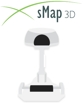
SMap 3D
heThis is our newest piece of technology which we purchased in late 2019. The sMap is revolutionary, prior to this machine we were only able to get half the information on the shape of the front of the eye.
Standard instruments measure the eye out to 11mm when we were using lenses that were 16.5mm. With the SMap instrument we can measure the shape of the white of the eye, which is where the lenses rest.
Many of our patients have BOTH corneal and scleral astigmatism, so we need to measure both! Having a lens that can correct the astigmatism in the cornea along with the sclera means the lenses are more stable, giving better safety and comfort for our patients with scleral lenses.